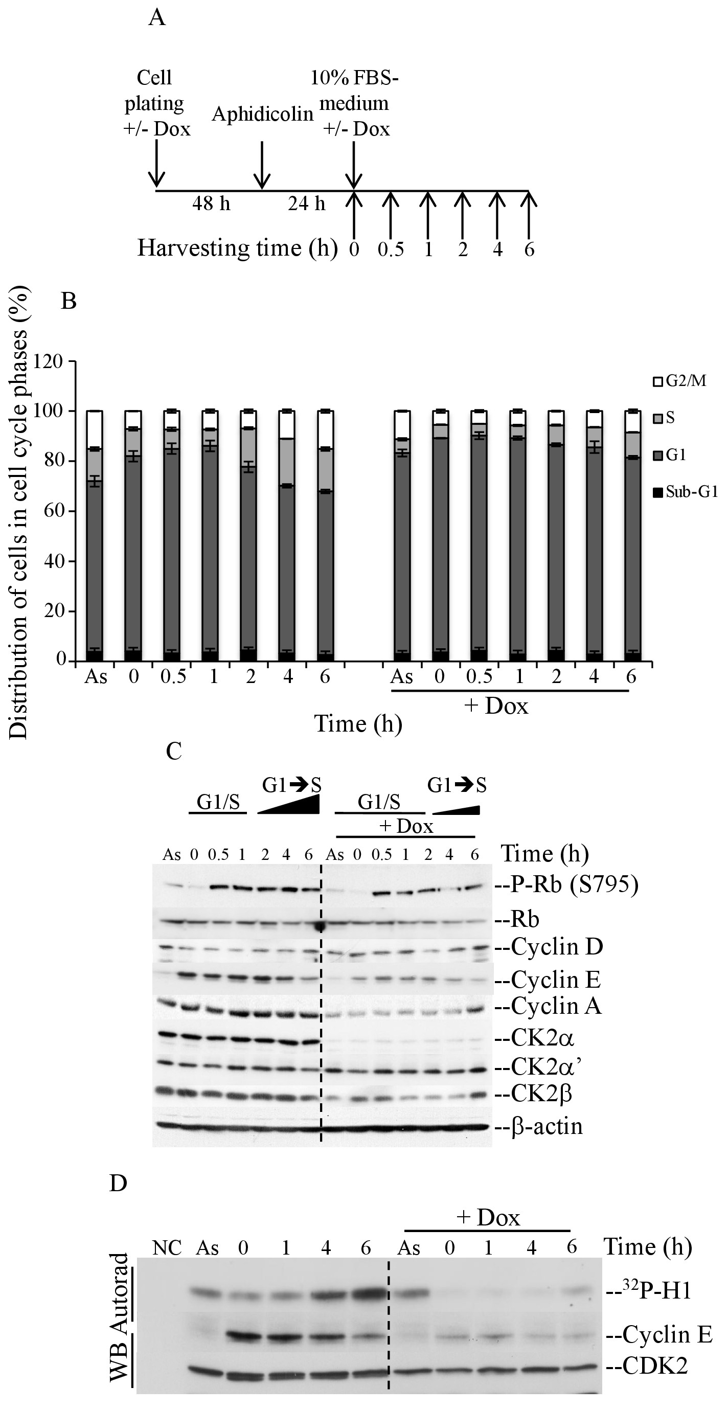
Fig. 3. Release from aphidicolin block causes delay G1/S cell cycle transition in cells depleted of CK2α. (A, B) Cells were synchronized in the presence of 3 μM aphidicolin for 24 hours as indicated in (A). After release from aphidicolin block, cells were harvested at the indicated time points and analyzed by flow cytometry (B). (C) Whole cell lysate from cells treated as reported in (A) was examined essentially as described in Fig. 2C. (D) Whole cell lysate was also employed for immunoprecipitation experiments with antibody against CDK2 or in the presence of control serum (NC). Cyclin E-CDK2 activity was measured by H1-based kinase assay. CDK2 and cyclin E protein levels were measured by western blot analysis using antibodies reacting against the indicated proteins. As: asynchronous cells.